Select or drop a image or 3D model here to search.
We support JPG, JPEG, PNG, GIF, WEBP, GLB, OBJ, STL, FBX. More formats will be added in the future.
Asset Overview
**Tricupsid Atresia**
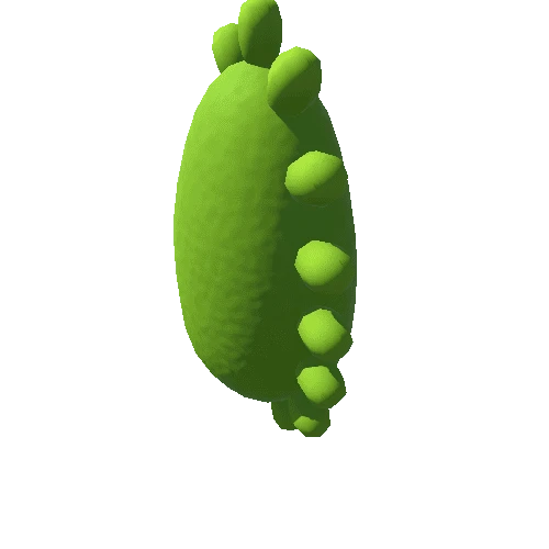
Multiple cardiac malformations, including muscular tricuspid valve atresia represented as a dimple in the floor of the right atrium. Minute right ventricular cavity (presumed secondary to large ventricular septal defect) primarily characterized in the conus region. In addition, the conus showed significant pulmonary infundibular stenosis and a bicuspid pulmonic valve. The left pulmonic artery arises lower than the right, slightly positioned to the right and is occluded (non-probe patent). A patch is attached to the external surface still not proble patent. There was a large unobstructed network of Chiari in the right atrium. The left atrium and two unusual appendages which were not thrombosed. The ventricular septal defect measured approximately 0.5cm in diameter; this matching the largest single dimension of the right ventricular chamber (also 0.5cm). the previously created atrial septal defect was still patent, measure 0.7 x 0.3cm with non-frayed and healed edges.


