Select or drop a image or 3D model here to search.
We support JPG, JPEG, PNG, GIF, WEBP, GLB, OBJ, STL, FBX. More formats will be added in the future.
Asset Overview
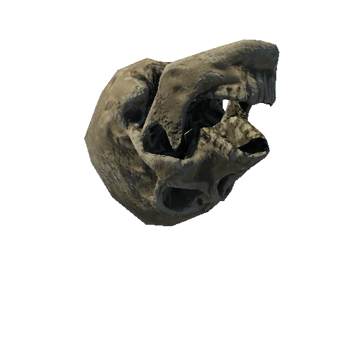
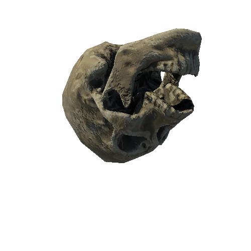
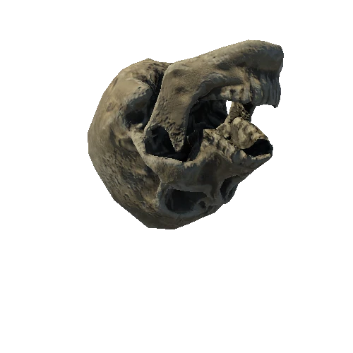
This is a plastinated human heart that was donated for research. The donor was an 89 year old female with a history of seizure disorders and Diabetes Mellitus. This model highlights a classic "four valve" view common in many medical textbooks. Here, the mitral and aortic valves are closed, and the pulmonary and tricuspid valves are open. Some trabeculae carnae can be seen in the Right Atrium through the tricuspid valve. An application of 3D scanning spray was used to facilitate the image capture. This heart uses reference ID: Heart0064.
We partner with [LifeSource](https://www.life-source.org/) to receive donated organs deemed not viable for transplant.
To learn more about cardiac anatomy, visit the [Atlas of Human Cardiac Anatomy](http://www.vhlab.umn.edu/atlas/index.shtml).
